
Ultrasound is a medical imaging procedure that employs high-frequency sound waves to produce pictures of the inside of the body. It is frequently used to see the heart, abdomen, pelvis, arteries, and other tissues. Additionally, ultrasound is used to monitor a fetus’ growth throughout pregnancy and to direct treatments like biopsies. The images produced by ultrasound are used to evaluate the health and structure of organs and tissues, and to guide medical procedures.
Ultrasonography has grown to be an essential tool for the diagnosis and management of a wide range of medical disorders. It does not use ionizing radiation and is non-invasive, safe, and reasonably priced, making it appropriate for repeated usage.
There are several different types of ultrasound, each with its own specific uses and applications. Some of the most common types of ultrasound include:
| Type | Description |
| Abdominal ultrasound | This type of ultrasound is used to create images of the organs inside the abdomen, such as the liver, pancreas, and kidneys. |
| Obstetric ultrasound | This type of ultrasound is used to evaluate the development of a fetus during pregnancy. |
| Pelvic ultrasound | This type of ultrasound is used to create images of the female reproductive organs and the urinary bladder. |
| Transvaginal ultrasound | This type of ultrasound is similar to pelvic ultrasound, but the transducer is inserted into the vagina to obtain images of the pelvic organs. |
| Transrectal ultrasound | This type of ultrasound is used to create images of the prostate gland and surrounding structures. |
| Cardiac ultrasound | This type of ultrasound is used to evaluate the structure and function of the heart and its blood vessels. |
| Vascular ultrasound | This type of ultrasound is used to evaluate the blood vessels, including the carotid artery, aorta and peripheral vessels. |
| Musculoskeletal ultrasound | This type of ultrasound is used to create images of the muscles, tendons, ligaments and joints. |
| Ophthalmic ultrasound | This type of ultrasound is used to create images of the eye and the surrounding structures. |
| Doppler ultrasound | This type of ultrasound uses the Doppler effect to measure blood flow through vessels. |
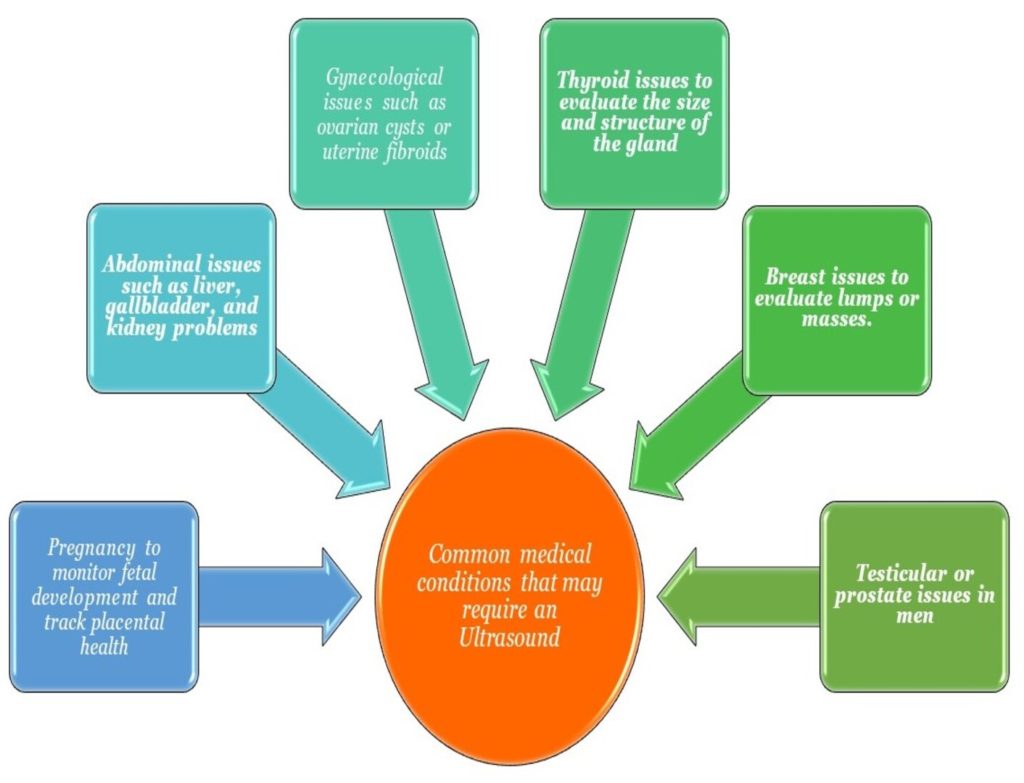
Some common medical conditions that may require an ultrasound for diagnosis include:
Preparation for an ultrasound may vary depending on the specific type of ultrasound and the area of the body being examined, but some common preparations include:
The procedure for an ultrasound typically involves the following steps:
The ultrasound results typically show images of the internal organs and tissues, including the liver, spleen, kidneys, bladder, uterus, ovaries, and others. The images produced by this test help doctors to diagnose various conditions, such as tumors, cysts, stones, and others. They can also help to monitor the growth and development of fetuses during pregnancy. Overall, the results provide important diagnostic information for healthcare professionals.
There are some benefits of Ultrasound:
Ultrasound is the first choice for diagnosis and a monitoring tool in present-day medicine.
Despite being regarded as a risk-free and non-invasive therapy, ultrasound has several possible drawbacks. The risk of receiving erroneous results depends on the patient’s weight and whether the area being checked is covered in bone or air. Certain illnesses, such as some forms of cancer or internal bleeding, cannot be detected by ultrasound.
Additionally, it is not advised for first-trimester pregnant women. If high-intensity ultrasound is utilized for an extended period, there is a slight chance that the tissue can become heated or produce bubbles (cavitation). It’s crucial to remember that other imaging modalities, such as CT or MRI, may be preferable for some disorders.
In conclusion, Ultrasound is a widely used diagnostic tool in medicine, providing valuable information about the internal structure and functioning of the body without the need for invasive procedures. In addition to the applications mentioned above, ultrasound is also used in fields such as physical therapy and sports medicine to help assess and monitor musculoskeletal conditions.
Moreover, advances in technology have allowed for the use of portable and handheld ultrasound devices, providing a convenient and accessible option for clinicians in various settings. With its versatility, safety, and ease of use, Ultrasound remains an important tool in the medical field.
Despite its many advantages, it also has certain drawbacks and risks, including the inability to detect some cancers or internal bleeding, and the possibility that alternative imaging modalities, such as CT or MRI, are preferable for specific situations. To decide the appropriate course of action, readers must share any queries or worries they may have with their healthcare professionals.
Ultrasound uses high-frequency sound waves to produce images of the inside of the body, while X-ray and CT scans use ionizing radiation. Ultrasound is non-invasive and does not use radiation making it safe for repeated use.
Ultrasound is generally considered a safe and non-invasive procedure with few risks. However, there are some risks and limitations associated with ultrasound that should be discussed with your healthcare provider.
Ultrasound is commonly used for a variety of purposes such as examination of internal organs, guidance for certain procedures, monitoring the development of a fetus during pregnancy, and evaluating the structure and function of the heart and blood vessels.
The procedure may take from 15 minutes to an hour, depending on the type of ultrasound and the area of the body being examined.
The risks include the potential for inaccurate results if the patient is obese, or if the area being examined is covered in bone or air. Ultrasound can’t detect certain conditions such as certain types of cancer or internal bleeding. Also, it’s not recommended for pregnant women during the first trimester.
You will feel a slight pressure when the transducer is pressed against your skin, but the procedure is generally painless.
In general, ultrasound can detect gallstones, kidney stones, liver problems, tumors, cysts, thyroid disorders, reproductive system issues, and monitor fetal health during pregnancy.
Usually for abdominal ultrasounds, only a light 6-8 hour fast is required and it is always better to avoid heavy or fatty meals. Sometimes, drinking water is required for bladder imaging.
Ultrasound is a diagnostic tool that can visualize and provide real-time images of internal organs and tissues; it can also aid in treatment, as well as in the monitoring of medical conditions, through image guidance.
The most common types of pregnancy ultrasound are early pregnancy scan, dating scan, NT scan, anomaly scan (Level 2), and 3D/4D ultrasound.
A Discount Providing Platform on all Diagnostic Services at your nearest Labs. 100's of Certified Labs, Assured Quality & Great Discounts.
