
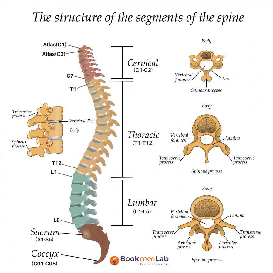
An MRI of the dorsal spine (also known as the thoracic spine) is an imaging test used to visualize the thoracic region of the spine. The thoracic spine is the middle part of the spine and runs from the base of the neck to the lower back. This test can identify abnormalities or injuries in the spinal cord, bones, and surrounding soft tissues. Some common conditions that it can diagnose include herniated discs, spinal tumors, and degenerative diseases such as osteoarthritis.
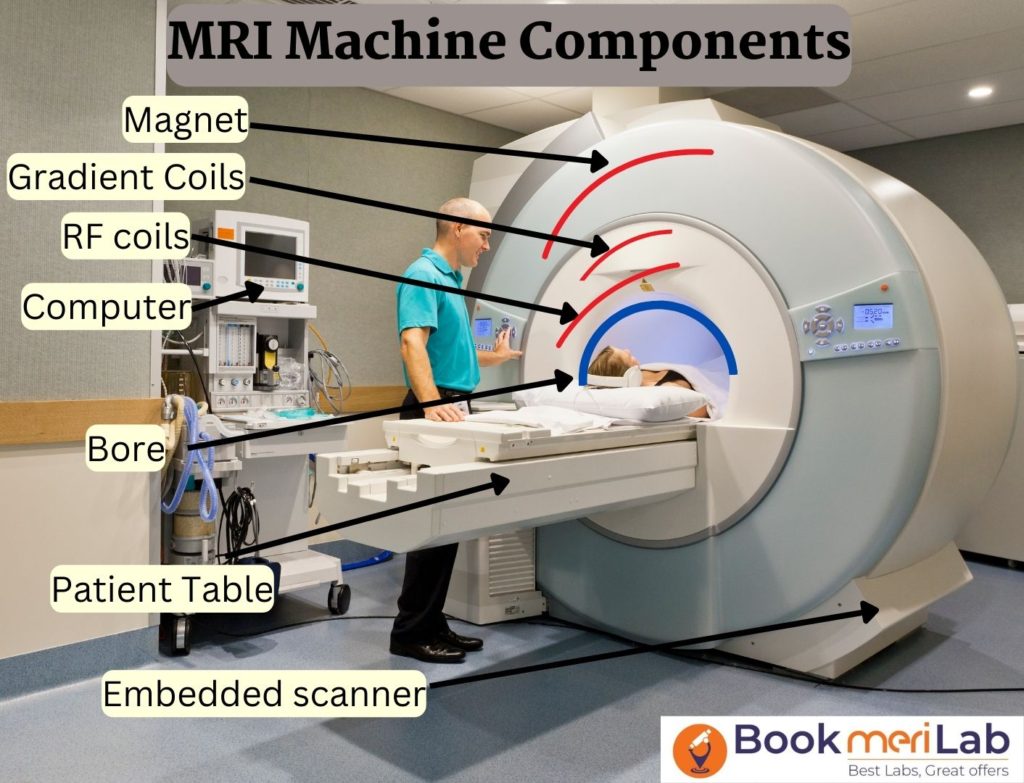
This test is generally safe, and painless and does not involve any ionizing radiations like X-Rays. It uses a strong magnetic field, radio waves, and a computer. The cost of an MRI Dorsal Spine ranges from ₹ 2700 to ₹ 8000.
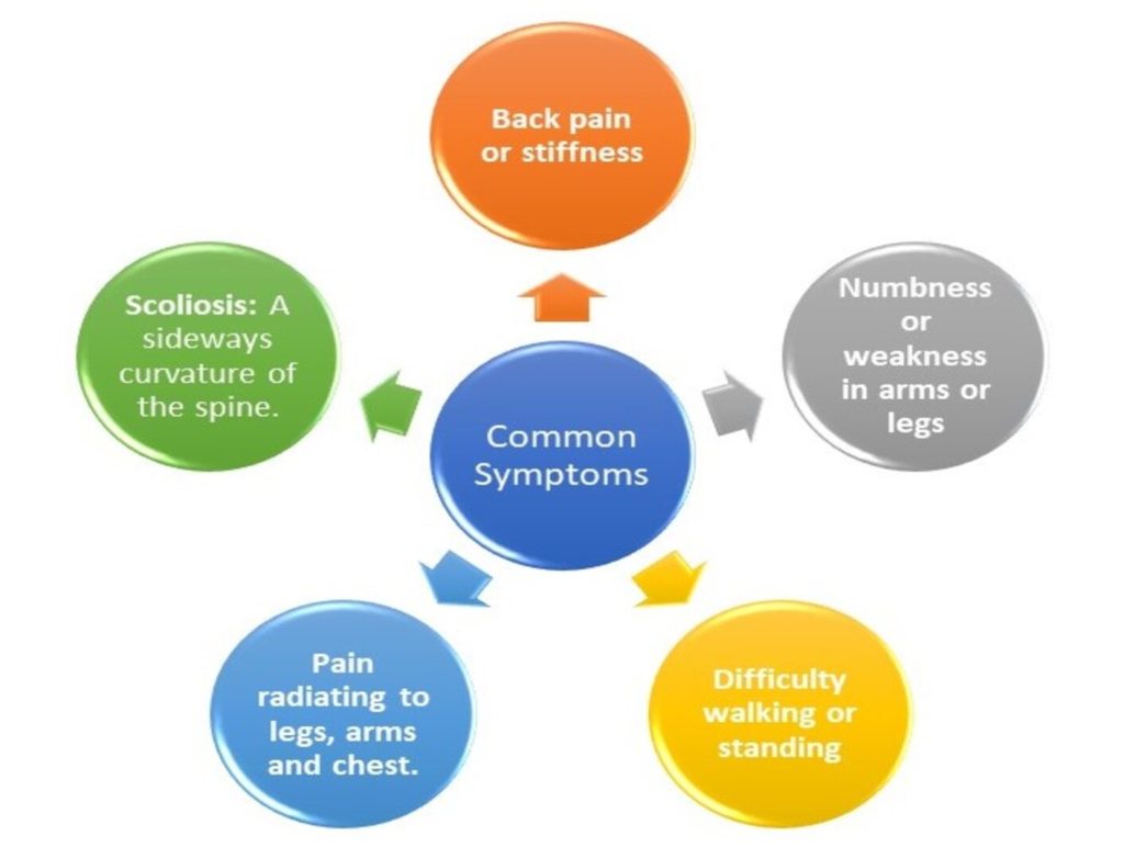
MRI Dorsal Spine is the procedure that solves the problems of back pain, numbness, tingling or limited mobility, if these problems have been going on for a long time. Detailed images of vertebrae, spinal cord, intervertebral discs, and even the soft tissues around the spine are provided by the MRI, which allows physicians to see the abnormal areas that are not visible in X-rays or CT scans. This, in turn, enables them to make the diagnosis at the earliest stage and to decide the proper treatment. The process is safe and non-invasive for the majority of patients, even those who need several scans. There is always the option of using a contrast material in some instances to make a certain area stand out and to facilitate the image. By selecting a trustworthy diagnostic center, one gets accurate results, reports on time, and the advice of a specialist for further treatment. Talking about the process and the cost of MRI Dorsal Spine with your doctor can be very helpful to the patients in their planning.
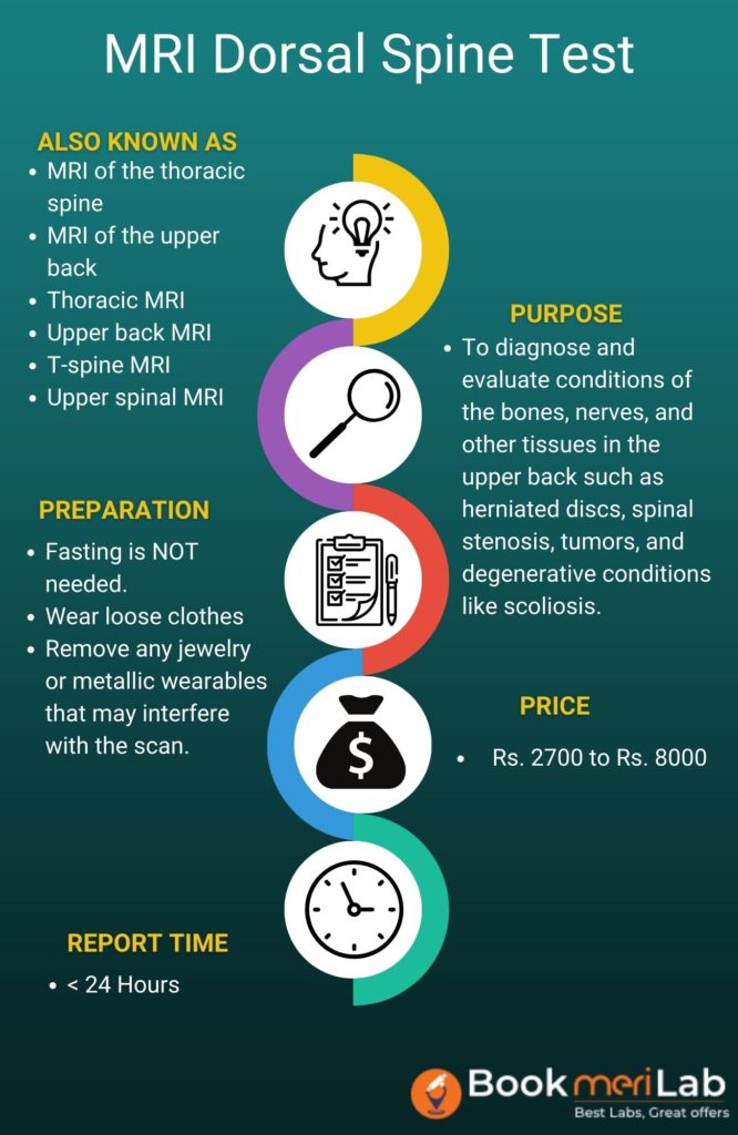
| Also Known As | MRI of the thoracic spine, MRI of the upper back, Thoracic MRI, Upper back MRI, T-spine MRI, Upper spinal MRI |
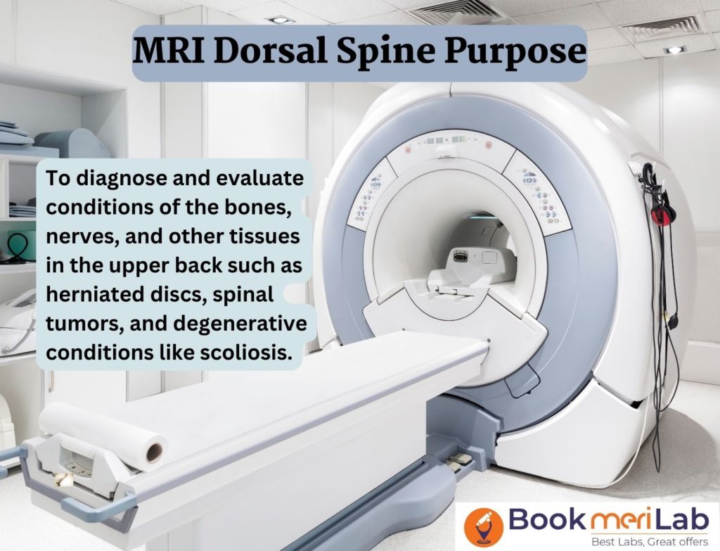
| Purpose | To diagnose and evaluate conditions of the bones, nerves, and other tissues in the upper back such as herniated discs, spinal stenosis, tumors, and degenerative conditions like scoliosis. |
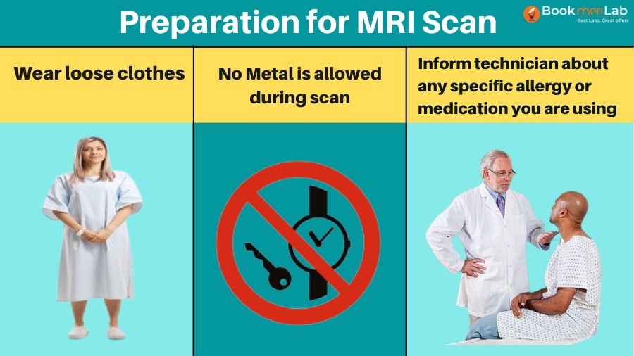
| Preparation | No special preparation is required. Remove any metal objects and wear comfortable clothing without metal parts. |
| Fasting | No |
| Reporting Time | <24 hours |
| Cost | ₹ 2700 to ₹ 8000 |
The cost usually ranges from Rs 2700 to Rs 8000. However, it may go up in cases involving special scans or if the test involves the use of a contrast media. The actual price would depend on the type of study, MRI centre, city, and technology (1.5 or 3 Tesla). Bookmerilab partner with more than 80 MRI centres in India and provide up to 50% discount. Contact us for more details.
The prices at some of the Popular MRI centres in India are listed below. 10-50% discounts are available at the below prices.
| Diagnostic Centres | Price |
|---|---|
| National Imaging | ₹ 2700 |
| Ganesh Diagnostics | ₹ 4800 |
| Mahajan Imaging | ₹ 6800 – ₹ 7200 |
| Aarthi Scans | ₹ 3285 |
| Healthmap Gurugram | ₹ 3307 |
| Srivastava MRI-NOIDA | ₹ 5250 |
| PULSE IMAGING | ₹ 4550 |
| Chhatrapati Shivaji Maharaj Hospital – KRSNAA | ₹ 2625 |
| NM Medical | ₹ 7225 |
| Shree Krishna Diagnostics | ₹ 4900 |
| Vijaya Diagnostic Centre | ₹ 7500 |
BookmeriLab has partnered with more than 80 MRI centres across India. We offer this test at an affordable starting price of Rs 2700 to Rs 8000. The table below shows the cost in major cities.
| Cities | Test Price Range |
|---|---|
| MRI Dorsal Spine Test Price in Delhi | ₹ 2700 – ₹ 7200 |
| MRI Dorsal Spine Test Price in Gurgaon | ₹ 3285 – ₹ 7200 |
| MRI Dorsal Spine Test Price in Noida | ₹ 2700 – ₹ 8000 |
| MRI Dorsal Spine Test Price in Mumbai | ₹ 2625 – ₹ 7225 |
| MRI Dorsal Spine Test Price in Kolkata | ₹ 3150 – ₹ 6650 |
| MRI Dorsal Spine Test Price in Bangalore | ₹ 2700 – ₹ 8000 |
| MRI Dorsal Spine Test Price in Hyderabad | ₹ 3285 – ₹ 7500 |
| MRI Dorsal Spine Test Price in Chennai | ₹ 3150 – ₹ 3285 |
This test helps diagnose and evaluate conditions of the upper and middle back region. Below we have listed the main purposes-
There are some benefits of MRI Dorsal Spine:
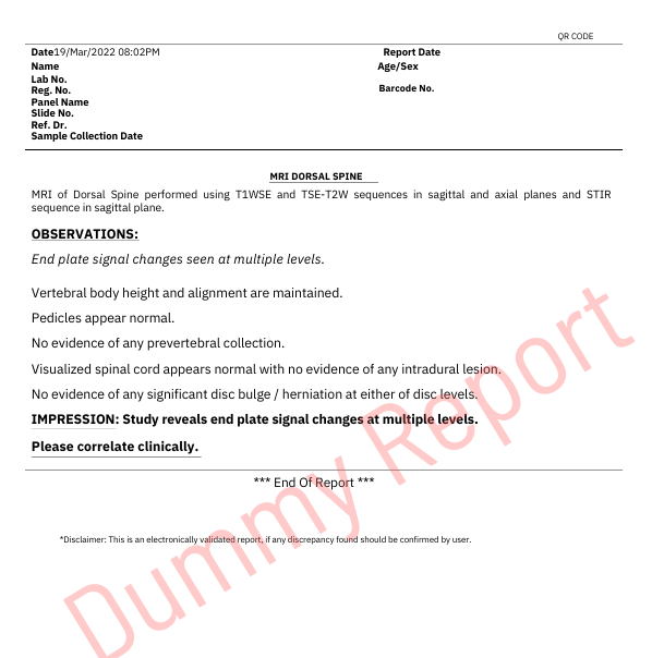
In a normal MRI scan of the dorsal spine, the vertebral bodies, intervertebral discs, spinal cord, and nerve roots should all appear to be intact and free from any problems.
Abnormal results may include:
The radiologist will provide the final interpretation of the MRI scan report and will take into account the patient’s clinical history, symptoms, and physical examination before reaching a diagnosis.
There may be some risks associated with an MRI such as-
In conclusion, an MRI of the dorsal spine produces accurate images of the spine’s soft tissues and bones without causing any pain. It can identify a wide range of spine problems as well as degenerative diseases. We can examine the dorsal spine with other imaging procedures such as X-rays, CT scans, and ultrasounds. However, MRI is the best option for the imaging of the soft tissues of the spine like intervertebral discs, nerves, etc. Before reaching a diagnosis, a radiologist will typically evaluate and interpret the results while taking the patient’s clinical history, symptoms, and physical examination into account.
In order to get a reliable diagnostic center for an MRI Dorsal Spine that will provide accurate results and reports on time, you have to make a wise choice. The method is safe, non-invasive, hence it is a good choice for most patients, even those who need multiple scans. There is a possibility of using contrast dye to make a particular area stand out and to give more detailed images. Preparation, procedure, and MRI Dorsal Spine price when discussed with the doctor will make the planning easy and effective.
By the union of sophisticated imaging technology and professional interpretation, the MRI Dorsal Spine is given a prominent place in the early detection, accurate diagnosis, and effective treatment planning, which, in turn, leads to the improvement of patient outcomes and spinal health.
A: An MRI of the dorsal spine is used to evaluate the bones, disks, and other structures of the upper and middle back.
A: The scan itself typically takes between 30 and 45 minutes.
A: You should wear comfortable, loose-fitting clothing and avoid wearing anything that contains metal.
A: No, an MRI is not painful.
A: You can eat and drink normally before an MRI of the dorsal spine.
A: No, there are no side effects associated with an MRI.
A: Yes, you can return to your normal activities immediately after an MRI.

Dr Garima Saroj is a dentist from Manipal college of dental sciences. She has previously worked at ESIC dental college and hospital Delhi and Dentecare multispecialty dental clinic Gurugram. She has a passion for writing for patient education and awareness.
A Discount Providing Platform on all Diagnostic Services at your nearest Labs. 100's of Certified Labs, Assured Quality & Great Discounts.
