
A mammography Test is an X-ray imaging technique that provides an internal image of the breasts. The test detects breast cancer and assesses the breast tissue’s size, shape, and density. It plays a significant role in detecting/ diagnosis the following:
The test uses a low dose of x-rays to diagnose any abnormality. During a mammogram, the breast is compressed between two plates to flatten it and spread the tissue out evenly. This allows the X-ray to penetrate the tissue more effectively and produces a clearer image. The test usually costs around Rs 700 – Rs 1800.
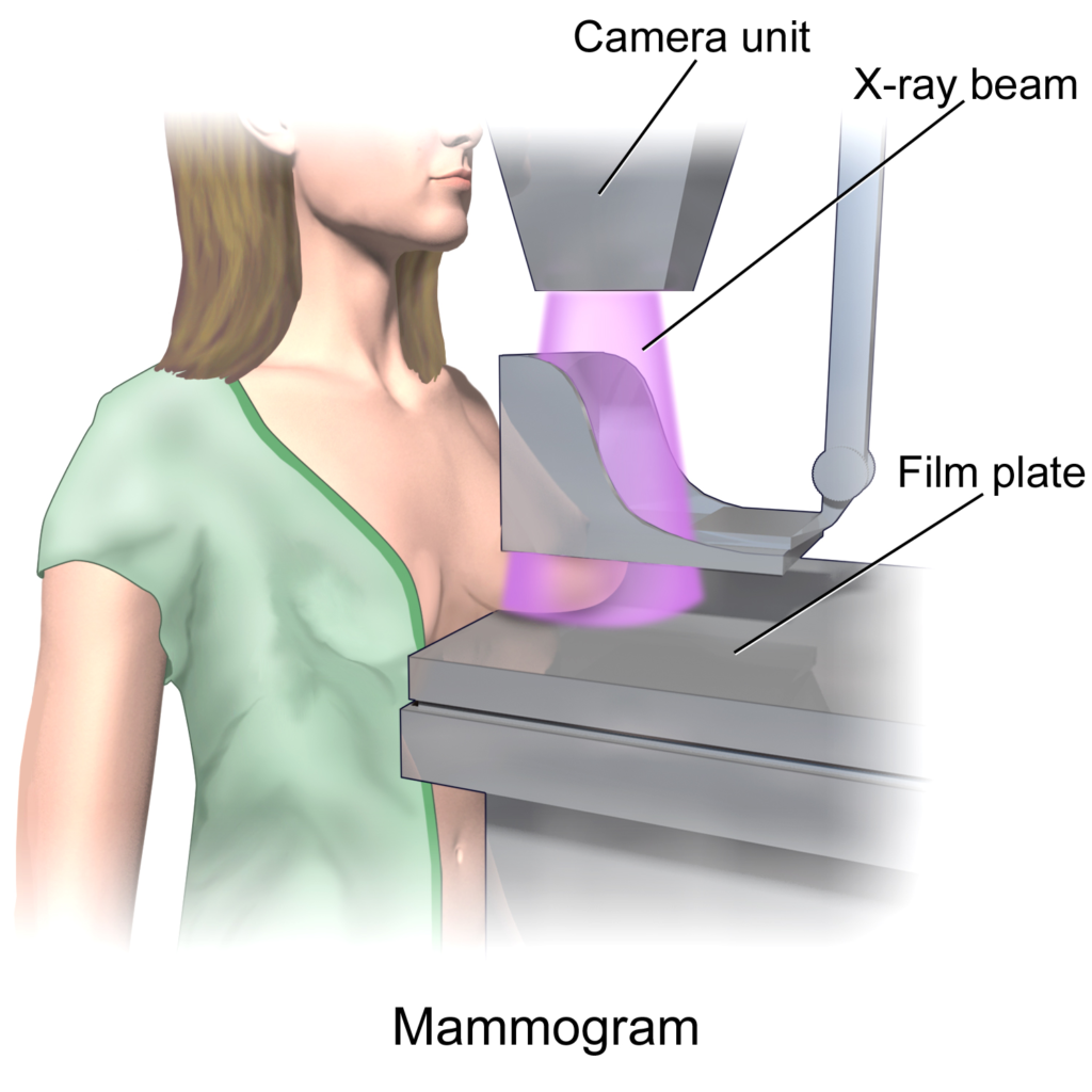
The X-ray image is then examined by a doctor or radiologist, who looks for any abnormalities or abnormalities in the breast tissue, such as lumps or masses that may be cancerous. If an abnormality is detected, the doctor may recommend further testing, such as a biopsy, to confirm the diagnosis.
| Also Known As | Mammogram, X-ray mammography, Sono-Mammogram |
| Purpose | Screening for Breast Cancer |
| Preparation | No special preparation required |
| Fasting | No |
| Machine Used | Mammography Machine |
| Reporting Time | 2 to 12 Hrs |
| Cost | Rs. 1500 to Rs. 3000 |
The cost of a mammography test in India typically ranges between Rs 1500 and Rs 3000. The price of a mammography test can vary depending on factors such as the city, location of the test, single or both breast scans and the type of mammography machine used.
The table below lists the prices at some of the popular diagnostic centres in India.
| Name of Lab | Test Price |
|---|---|
| Aarthi Scans | Rs 1800 |
| Janta X-Ray | Rs 650 |
| Dr Lal Pathlabs | Not Available |
| Mahajan Imaging | Rs 1800 |
| Vijaya Diagnostics | Rs 1500 |
| Pulse Imaging | Rs 1050 |
| NM Medical | Rs 1275 |
| Medivision | Rs 1173 |
The Mammography test cost depends upon the one recommended by your healthcare professional. The cost varies depending on the lab and the city. The table below list the price for single breast mammogram.
| City | Test Price |
|---|---|
| Mammography Price in Delhi | Rs 650 to Rs 1800 |
| Mammography Price in Mumbai | Rs 750 to Rs 1800 |
| Mammography Price in Bangalore | Rs 1800 |
| Mammography Price in Hyderabad | Rs 750 to Rs 1800 |
| Mammography Price in Gurgaon | Rs 1200 to Rs 1800 |
| Mammography Price in Chennai | Rs 1100 to Rs 1800 |
| Mammography Price in Kolkata | Rs 1800 |
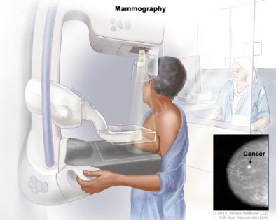
In Mammography, a woman’s breast is scanned under an X-ray dose. In this test, a small X-ray dose is exposed to the breast. This is painless and non-invasive (that involves no cuts and incisions). The scanned images of the internal structure of the breasts give a detailed analysis of the internal structure of the breasts.
There are 5 types of Mammography.
Sono mammography uses the ultrasound technique for the diagnosis of any structural abnormality in the breast. But x-ray mammography involves the use of X-rays.
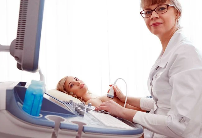
Doctors may recommend mammography tests for symptoms such as the presence of a lump in the breast, pain, or nipple discharge.
1. Screening: The test can detect abnormal changes in the breast before the patient or doctor gets to know it. Women over 50 years and above are recommended to get a mammography screening yearly. Doctors may recommend other tests along with mammography screening. This depends on risk factors such as family history, ovarian cancer, etc.
2. Diagnosis: Your doctor may recommend Mammography to diagnose abnormal findings during physical examination.
3. Breast cancer: A widely used test to diagnose cancer or tumour’s early development and stages.
4. Fibroadenoma: Fibroadenoma is a medical condition in women that causes non-cancerous lumps in the breast. The non-cancerous lumps are not dangerous and cause no disease.
X-rays are used in Mammography to get an internal image of the breast. The mammography equipment emits radiation on the breast, which is recorded in a film.
Step 1- The female technician will instruct you to place your breast on a plastic platform. Another plastic plate compresses the breast. The plastic plate is held still while an x-ray is taken.
Step 2- Mammography positioning: You change position per your technician’s direction. This helps in getting images from different angles.
Step 3- You may be asked to remain still and hold your breath while taking the x-ray. The quality of the scanned picture relates to the stillness of the breast.
Some women find having a mammogram uncomfortable due to factors like the size of their breasts and how much they need to be pressed. Breasts get tender when you are about have your period. Hence they can get sensitive. But a skilful technologist will take care of these things.
You should consider the following points:
Theoretically speaking, Excessive radiation exposure can cause cancer. However, radiologist makes sure that the lowest amount of radiation is used while producing the best possible image. The latest technology in mammography equipment produces high-quality images with a low dose of radiation. So, the advantages of mammography tests outweigh the risks associated with them.
Test results are usually available within 12 hours. Most diagnostic centres provide results immediately after the scan.
If the result is normal, make sure you get the test done annually. This helps the doctors compare the image and look for any changes.
The normal result indicates the absence of abnormal cancerous and non-cancerous lumps in the breast. The White area in the mammograph images represents the healthy tissues.
Abnormal results may require further other tests to confirm the diagnosis. Your doctor will proceed with follow-up exams to look for the presence or absence of cancer.
The radiologists use the scoring system ‘BI-RADS’ to represent mammography results.
The main types of abnormalities found in the mammogram are
BI-RADS is the Breast Imaging Reporting and Database System score. The score is categorized from 0 to 6. The score helps your doctor determine the line of treatment and monitor the treatment progress. Follow what your healthcare provider recommends, as early diagnosis can treat your breast cancer.
The table given below describes the assessment of BI-RADS as per the category.
| SCORES | SIGNIFICANCE |
|---|---|
| 0 | Incomplete test and requires additional tests. |
| 1 | A negative result (No cancer) |
| 2 | Normal results (but the presence of benign cysts) |
| 3 | Probable normal results (with 2% chance of cancer). Follow up required |
| 4 | Abnormal results (20-35% possibility of cancer). 4A-Low suspicion of cancer 4B-Moderate suspicion of cancer 4C-High suspicion of cancer |
| 5 | The abnormal result (95% chance of breast cancer). |
| 6 | After a biopsy of breast cancer is confirmed. |
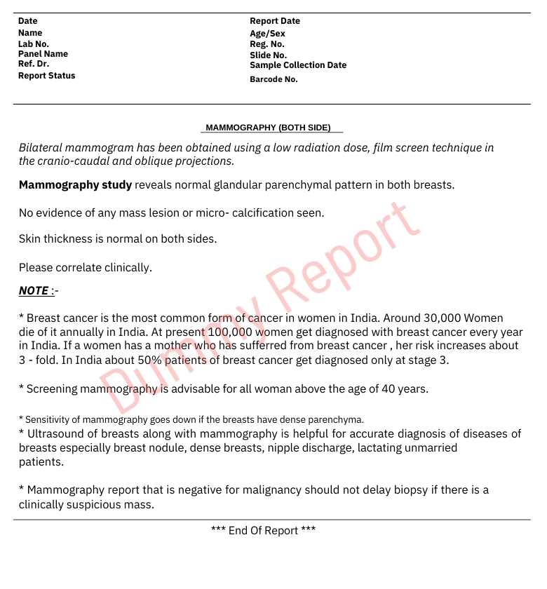
Here is the actual report of mammography would look like:
Mammography is an easy but very effective test that has a very big influence on the health of women. This test is used to identify the presence of breast cancer and other breast-related problems, along with the latter stages of the disease, i.e., a stage in which there are no visible symptoms. Early detection through mammography is followed by timely therapy, which dramatically increases the survival rate. A few women might experience a little pain during the procedure, but the feeling of reassurance and preventive care, which is given to them, is much greater. Regular mammography screening, which is the doctors’ recommendation, is the step that plays a very big role in keeping the health of the breast and giving consolation to women of all age groups.
The entire procedure takes around 10- 20 minutes.
The high breast density may suggest that you have breast cancer.
The experience of Mammography varies. The procedure can be painful or cause no pain. Most women feel discomfort during the entire procedure.
The increased breast density may produce unclear mammography scans.
Even though doctors say that it’s rare to get breast cancer after 1 year of mammography, one should continue self-examination of breasts since it’s not impossible.
The USPSTF says that women having an age 50 or above are at the average risk of breast cancer and should get mammograms done after every 2 years. Women with age between 40 to 49 should consult a doctor and decide whether they should take the test.
Talking about mammography accuracy, about 6 – 8%, the test has produced false positives. But, on the dangerous side, about 17% of breast cancers are missed. Hence a clinical examination needs to be followed.
Well, breast implants can hinder your scanning, depending upon where they have been placed, especially if they are placed above the chest muscle.
Yes. The compression of the breast is essential because of the following points:
1. It spreads the breast tissues, and this increases the surface area. This increased surface area exposes even the tiny abnormalities in the breast.
2. It minimizes the blurring of the image as the part is held still. Therefore, the images produced would be clear.
3. A complete reduction in the scattering of x-ray scatter results in the sharpness of the image.
Mammography is a highly effective tool for detecting many types of breast cancer, but it may not catch all forms, especially in younger women or those with dense breast tissue. Regular screenings are still crucial for early detection.
Dr Bhumika Rana is a Bachelor of Ayurvedic Medicine & Surgery (B.A.M.S.). Earlier She was an Operation Specialist at AIIMS Delhi & a Medical officer with the National health mission.
A Discount Providing Platform on all Diagnostic Services at your nearest Labs. 100's of Certified Labs, Assured Quality & Great Discounts.
